Vision Disorders
Structure of the Eye
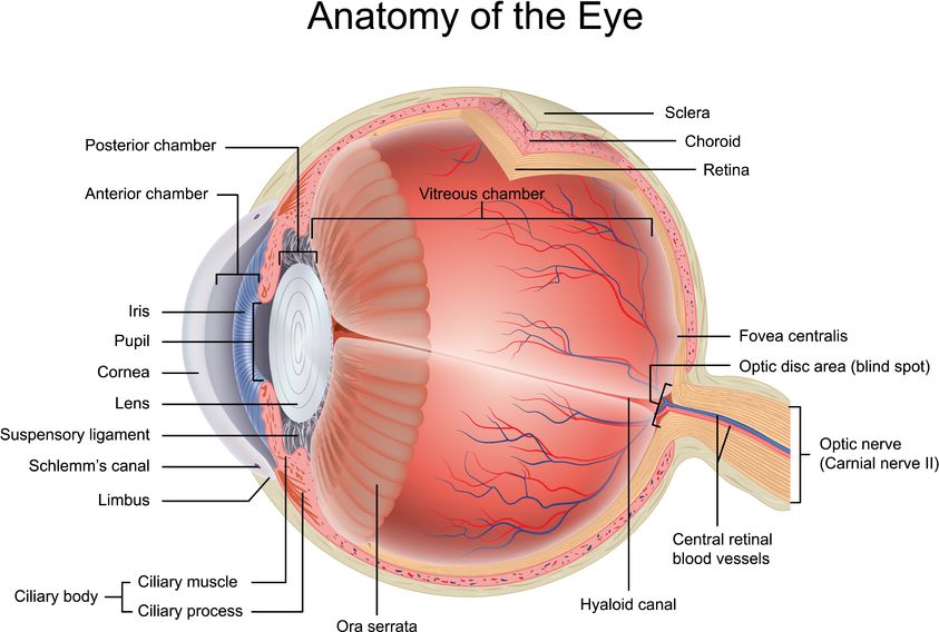
The eyeball has a diameter of about 24 mm. The eye works somewhat like a camera. The cornea, pupil, and lens create a sharp image of our surroundings on the retina. The lens changes shape to help focus light on the retina.
Cornea — The curved outer layer at the front of the eyeball. It helps focus incoming light. The cornea gets its nutrients from tears and fluid inside the eye. It has no blood vessels, and its focusing power is always the same.
Sclera — The white, protective outer layer of the eye. It keeps the eye safe from injuries and provides attachment points for the muscles that move the eyeball. The optic nerve and blood vessels pass through the back part of the sclera.
Iris — The colored part of the eye located behind the cornea. It has a hole in the center called the pupil. The iris contains muscles that adjust the size of the pupil. This helps regulate the amount of light entering the eye based on the surrounding light levels. It's an automatic process called light adaptation.
Lens — Located behind the iris, it is a transparent, oval structure. The lens further bends light that passes through the pupil. It can change shape to focus on objects that are close or far away. This ability is called the eye's accommodation.
Retina — The inner layer of the eyeball that senses light. It contains cells called rods and cones. Rods detect light intensity, while cones help us see colors.
Macula (fovea centralis) — A small area in the center of the retina. It has the highest concentration of cones and provides the clearest vision.
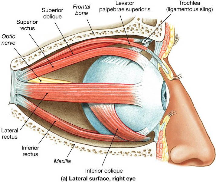
Eye muscles — Six muscles on the outside of the eye move the eyeball. There are four straight muscles (superior, inferior, medial, and lateral) and two oblique muscles. These muscles work together to control eye movements. In the Bates Method, their action is also considered to influence the length of the eyeball and the way we see.
Myopia (nearsightedness)
Myopia is one of the most common refractive errors and, by some estimates, affects nearly half of the world's population. It usually first appears as difficulty seeing distant objects clearly. In many people, myopia may progress over time, especially during adolescence; the rate varies from person to person.
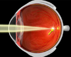
Anatomically, myopia usually involves a mismatch between the eye's focusing power and its length. In myopia, the power of the eye's optical system is too great or the eye is too long, resulting in a blurry image of distant objects. People with myopia usually see near objects more clearly than distant ones.
Myopia is typically corrected with diverging glasses or contact lenses. Their optical power is expressed in diopters with a minus sign. In cases of high myopia, degenerative changes in the choroid, retina, and vitreous body may occur later in life. Myopia most often develops during adolescence.
Hyperopia (farsightedness)
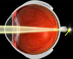
Farsightedness (also called hyperopia) is a refractive error caused by a disproportion between the power of the eye's optical system and the length of the eyeball. In hyperopia, the power of the eye's optical system is too weak or the eye is too short, which can blur near vision and sometimes distance vision as well.
Thanks to the ability of the eye's optical system to add power (accommodation), a person with hyperopia usually sees distant objects clearly, but experiences the most discomfort when working up close. Presbyopia is a separate age-related focusing change that can cause similar near-vision symptoms, as the lens becomes less flexible with age.
Hyperopia is typically corrected with corrective glasses or contact lenses. These are converging lenses. Their optical power is indicated in diopters, with a plus sign (e.g., plus 3 diopters).
Presbyopia (age-related farsightedness)
Presbyopia is a progressive decline in the eye's ability to change its focusing power. It is a natural part of eye aging and affects everyone, regardless of previous refractive errors. People aged 40–45 begin to experience difficulty reading, especially small print, typically marked by the need for brighter lighting and for holding text farther from the eyes.
Early presbyopia symptoms are more bothersome in the evening, when the eyes are tired. This progressive "aging" of the eye eventually leads to an inability to read without dedicated reading glasses. Presbyopia can be corrected with bifocal or progressive glasses.
Astigmatism (corneal irregularity)
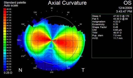
Astigmatism is a refractive condition that causes distorted vision due to the asymmetry of the eye's cornea. If the curvature of the cornea in the vertical plane is different from that in the horizontal plane, light rays hitting different parts of the cornea are refracted to varying degrees. This causes the image to appear blurry. When viewing a cross, some people see either the vertical or horizontal line more clearly than the other. This type of astigmatism is called regular, and such an eye has two focal points. To correct this condition, glasses with cylindrical lenses are used. The human eye usually has a minor natural irregularity of up to 0.5 D, which does not require correction. Eye injuries often cause an uneven corneal surface, resulting in irregular astigmatism, characterized by a greater number of focal points. To correct this condition, rigid (or specialized soft) contact lenses are required.
Eye Convergence Problems
When the eyes work together normally, they effortlessly focus on a given object by turning inward together. At short distances (e.g., reading a book), the eyes automatically turn inward to maintain a sharp image. At long distances, the eyes are virtually parallel. In a dark environment, where the pupils are wide open, the depth of field is minimal. In bright light, with small pupils, the depth of field is much larger, and we see much more sharply.
Thanks to eye convergence, we perceive depth and see the world in three dimensions. The brain automatically overlays the images from each eye, creating a single three-dimensional image. If you have problems with eye convergence, you may not be able to accurately judge the distance to objects. Convergence can become weaker over time for some people, often alongside presbyopia, which is why it's important to check for problems regularly and, if necessary, begin exercises that support eye-convergence practice. Some people find these exercises helpful, so if convergence problems affect you, it may be worth dedicating some time to them. More significant convergence problems can occur in people with strabismus.
Strabismus (crossed eyes)
Eye movement in every direction — which lets us observe both still and moving objects across the whole field of vision — depends on the proper functioning of the external eye muscles. Under normal conditions, both eyeballs are aligned in parallel. In a process called fusion, the visual cortex overlays and merges the two retinal images (from the right and left eye) into a single stereoscopic image.
Strabismus is an abnormal alignment of the two eyeballs. It stems from an imbalance of the eye muscles, accompanied by disturbed retinal correspondence or a lack of binocular vision. Strabismus can cause amblyopia (commonly called lazy eye) — reduced vision in one eye.
Strabismus is often classified by the direction of the eye deviation:
- When an eye turns inward, it's called convergent strabismus, or esotropia.
- When an eye turns outward, it's called divergent strabismus, or exotropia.
- When an eye turns upward, it's called hypertropia.
- When an eye turns downward, it's called hypotropia.
- When the eyes are misaligned diagonally, it's called oblique strabismus.
The most common form, accounting for almost half of strabismus cases, involves excessive tension in the medial rectus muscle, causing the eye to turn inward too much. The deviation can be minimal and almost unnoticeable, or so severe that the pupil nearly disappears into the corner of the eye. Because the deviation of one eye's axis leads to double vision, the brain suppresses the image from the misaligned eye, which results in reduced vision (lazy eye). For this reason, strabismus is usually associated with poorer vision in the deviating eye.
Latent (hidden) strabismus is often considered easier to work with, because it appears only when one eye is used alone; with both eyes open, binocular vision keeps the eyes converged and the strabismus does not show. The work here relies mainly on exercises that improve convergence and balance the refractive error between the eyes.
Strabismus often appears in children but also occurs in adults. Its exact causes are not always clear. Conventional treatment usually addresses only the reduced vision, if present. Strabismus can be corrected with prismatic glasses that compensate for the deviation, or surgically. However, these methods are often ineffective and can lead to mounting frustration for everyone involved. Some people use natural vision training as a complementary practice, and children often respond especially well to daily practice. Even so, if you suspect strabismus, especially in a child, do not rely on exercises alone: consult an ophthalmologist or optometrist for a proper evaluation.
Causes of strabismus:
- Strabismus can occur in healthy eyes without a specific cause.
- It can be caused by eye conditions that result in poorer vision in one eye.
- Some cases of strabismus are related to conditions that hinder the development of binocular vision, such as cataracts.
- Strabismus can also be associated with retinal changes that affect the proper perception of visual stimuli.