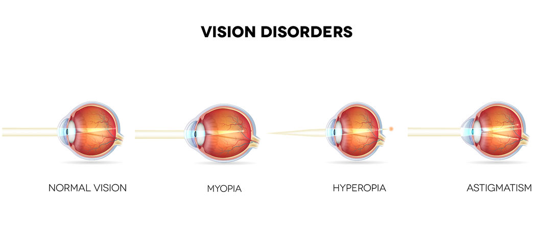
The eyeball is 24 mm wide. The eye acts in a similar way to a video camera. The cornea, pupil and lens generate a sharp picture of the surrounding view on the retina. The lens changes its shape automatically, and the corresponding muscles alter the size of the eyeball, which allows for generation of perfectly sharp picture on the retina.
Cornea – a convex external layer of the eyeball located in its front part. The cornea has a watery liquid behind, and further behind is the lens. The cornea has no vessels and is fed through the diffusion – by tears and watery liquid of the eyeball. The cornea focuses the light rays stronger (about 43 D) than the lens (about 20 D). However, unlike the lens, the cornea’s power to focus the rays is fixed (cannot be adjusted).
Sclera – protects the eye against mechanical damage. It has eye muscles attached that move the eyeball. In the rear side of the eye, the sclera is crossed by the optic nerve and blood vessels.
Iris - located on the front side of the eye, behind the cornea, is a circular membrane which has an aperture inside. This aperture, called the pupil, lets the sunrays enter the eye. The iris has smooth muscles that constrict or expand the pupil. This process happens involuntarily and is aimed at adjusting the amount of light that reaches the eye according to the light intensity of the environment. It is known as the adaptation of the eye.
Lens – located behind the iris, has a transparent, oval structure. Its function is to further refract the light rays crossing the pupils. The lens can change when we are looking at the objects located near or far from the eye. This ability is known as the accommodation of the eye.
Retina – the internal membrane that forms a light-sensitive layer of the eyeball. Its function is to receive the light sensations. Its elements responsible for this function are the light-sensitive cells – retinal rods and cones. The rods receive the sensation of light intensity, and the cones are used to differentiate between the colors.
Macula lutea – a spot on the retina, characterized by the best visual acuity, located on the visual axis. It has the densest concentration of cone cells, and no rod cells.
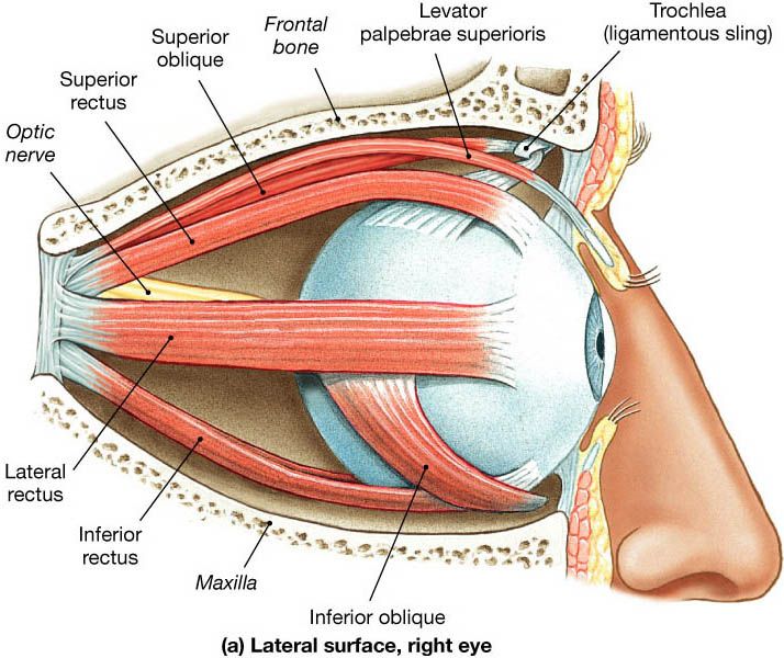
Eye muscles. The eyeball is put in motion by six external eye muscles. Four superior muscles: Four rectus muscles: superior, inferior, internal rectus muscle and external rectus muscle, which have their rear attachments far behind the eyeball. Much different and more complex is the system of oblique muscles which are responsible for the particular eye movements, as well as shortening and lengthening of the eyeball.